Have you ever performed an immunohistochemical (IHC) stain that showed a general “blush,” with only subtle differences between samples or experimental treatments? Semi-quantitative scoring methods like Allred may not reveal meaningful separation. Or perhaps you have a large collection of stained tissue sections, but not the time, personnel, or pathology resources required to score them consistently and confidently. If staining differences weren’t obvious, those slides may now be sitting on a shelf—archived, unused, and full of unanswered questions.
Today, that no longer has to be the end of the research.
Modern digital pathology tools make it possible to extract objective, quantitative data from archived IHC slides, breathing new life into past experiments and enabling deeper insight without repeating the original wet lab work.
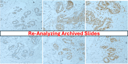
Re-Analyzing Archived IHC Slides with HALO® Image Analysis
Instead of relying on subjective visual scoring, HALO® applies consistent, standardized parameters across entire tissue sections to generate reproducible, quantitative outputs. Large batches of stained tissue sections can be analyzed using the same precise parameters, ensuring consistency across entire datasets. This approach allows researchers to revisit older studies and ask new questions—often revealing patterns that were difficult or impossible to resolve using manual scoring alone.
This approach allows researchers to revisit older studies and ask new questions—often revealing patterns that were difficult or impossible to resolve using manual scoring alone.
A Real Example: Re-Analyzing p53 IHC Slides from Archived Tissue
One example involves p53 immunohistochemical staining performed on human breast tissue samples originally generated in 2007. The original study asked whether reproductive history influenced tissue sensitivity to oxidative stress or radiation-induced p53 responses.
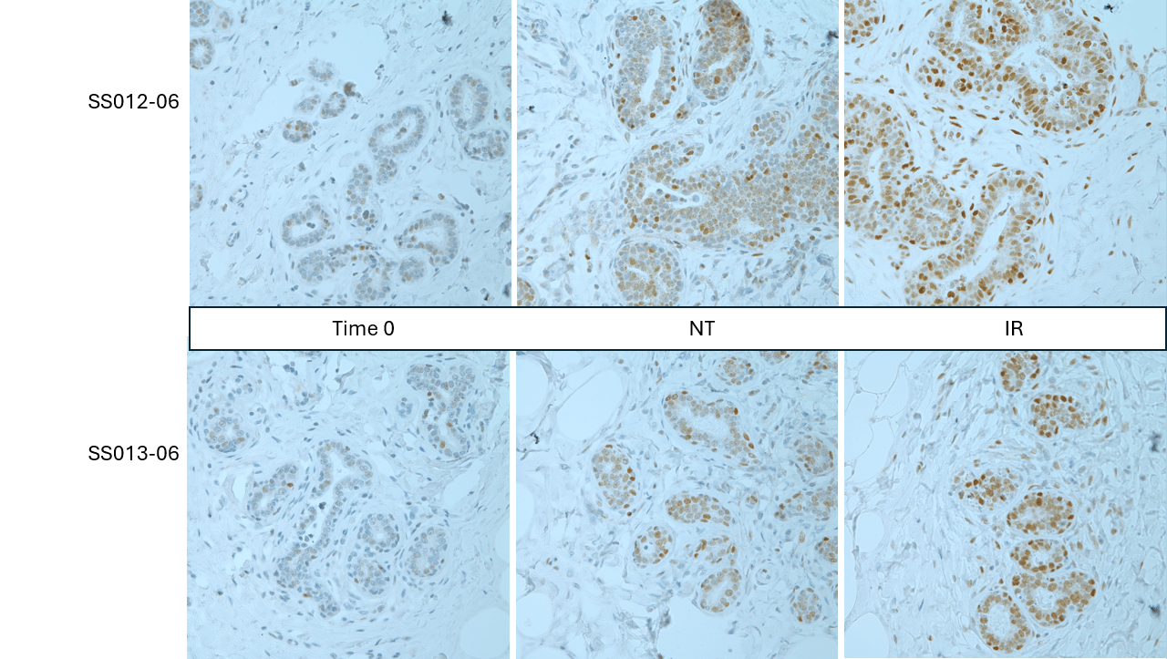
Tissues were collected and fixed immediately after surgery (time 0) or placed into explant culture conditions for several days, where they were either exposed to ionizing radiation (IR) or left untreated (NT). This experimental design resulted in biologically meaningful but often subtle differences in p53 staining.
At the time, slides were evaluated by two blinded reviewers using a semi-quantitative scoring approach:
⦁ Staining intensity: 0–3
⦁ Percent epithelium stained: 0–3
While this method captured overall trends, it offered limited dynamic range and introduced reviewer-to-reviewer variability.
To better understand how digital re-analysis expands dynamic range beyond traditional visual scoring, HALO®-generated H-scores were calculated from the same archived slides
Understanding the H-Score
The H-score (histochemical score) integrates both staining intensity and the proportion of positive cells within a tissue section. Staining intensity is categorized as negative (0), weak (1), moderate (2), or strong (3). The H-score is calculated as: H-score = (1 × % weak) + (2 × % moderate) + (3 × % strong)
(Range: 0–300)
|
Tissue ID and Treatment
|
Blinded Reviewer Score
|
HALO® H-Score
|
|---|---|---|
|
J05-30 time 0
|
1+2
|
2.9
|
|
J05-30 24 hrs 6 hrs NT
|
1+3
|
48.1
|
|
J05-30 24 hrs 6 hrs IR
|
2+3
|
50.7
|
|
SS0011-06 time 0
|
1+2
|
6.9
|
|
SS0011-06 24 hrs 6 hrs NT
|
2+2
|
47.9
|
|
SS0011-06 24 hrs 6 hrs IR
|
2+3
|
194.5
|
|
SS0013-06 time 0
|
1+2
|
8.6
|
|
SS0013-06 24 hrs 6 hrs NT
|
2+3
|
79.5
|
|
SS0013-06 24 hrs 6 hrs IR
|
2+3
|
147.2
|
|
SS0012-06 time 0
|
1+2
|
22.4
|
|
SS0012-06 24 hrs 6 hrs NT
|
1+3
|
66.9
|
|
SS0012-06 24 hrs 6 hrs IR
|
2+3
|
117.4
|
Figure: Manual intensity/percent scores generated in 2007 compared to H-scores obtained by re-analyzing the same archived IHC slides using HALO® image analysis.
Visualizing Donor-Level Trends with Quantitative Image Analysis
While the table above preserves the original scoring framework and allows direct comparison to historical methods, visualizing the HALO®-generated H-scores across conditions reveals the biological trends more clearly. When plotted by donor, p53 expression increased following explant culture in 4 of 4 donors, with a further increase following ionizing radiation in 3 of 4 donors– patterns that were difficult to appreciate using ordinal visual scoring alone.
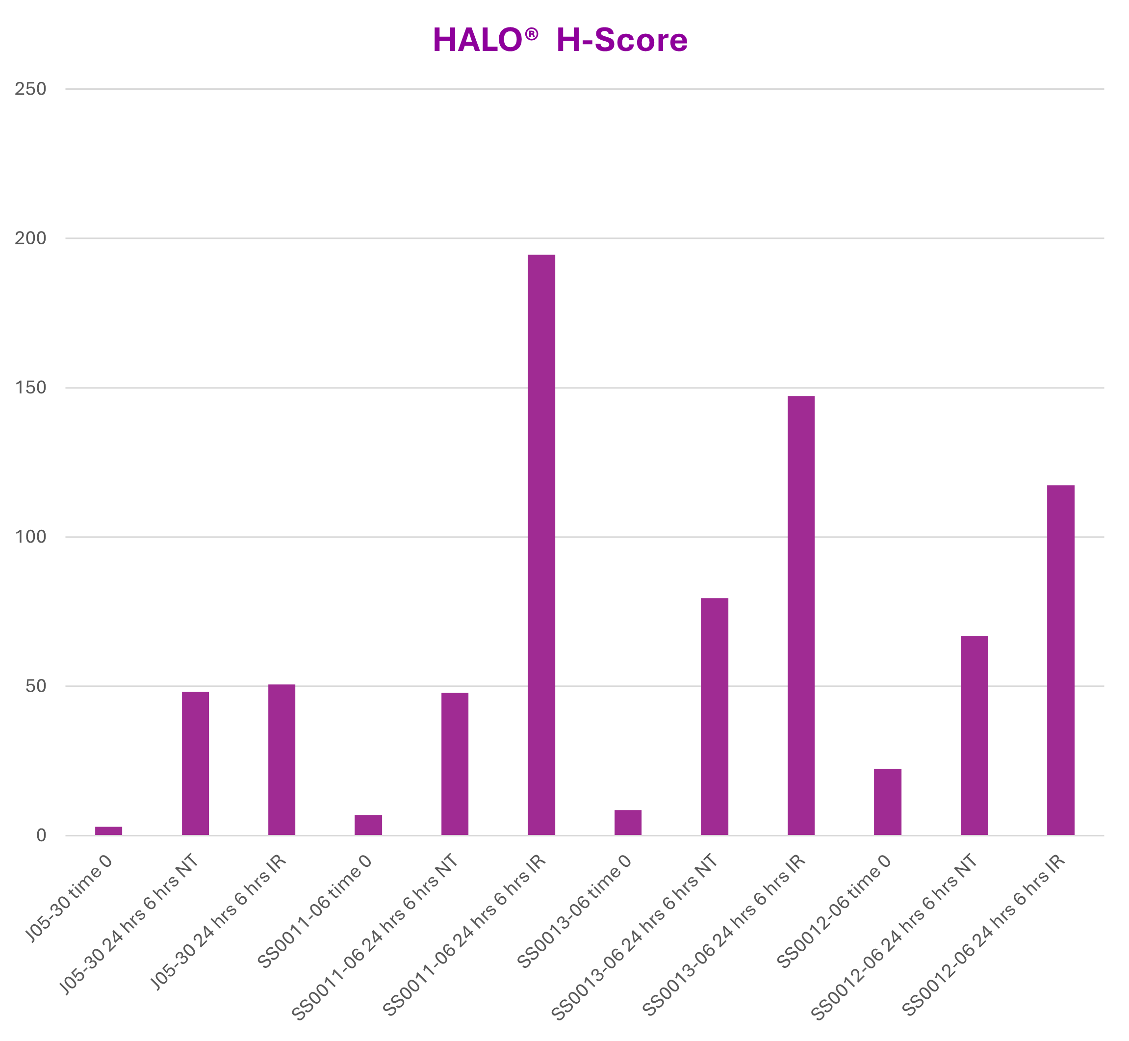
Figure: HALO®-generated H-scores for archived p53 IHC samples plotted by donor and treatment condition. Quantitative image analysis reveals consistent increases in p53 following explant culture and additional induction following ionizing radiation (IR) in most donors.
Interpreting the Impact of Digital Re-Analysis
While both approaches detected similar biological trends, HALO® image analysis revealed a substantially broader dynamic range (2.9 to 194.5 compared to 3-5 with traditional scoring) and removed the subjectivity inherent to visual scoring—particularly in cases where blinded reviewers did not fully agree. By applying consistent, standardized parameters across all samples, HALO® enabled reliable quantification of archived p53 IHC data, bringing new life to a study originally generated in 2007.
What Types of Archived IHC Slides Benefit Most?
To ensure reliable, reproducible results across large slide sets, HistoSpring applies a standardized digital pathology workflow designed for studies where traditional scoring methods fall short. This approach is particularly well suited for:
⦁ Slides with graded or subtle DAB staining patterns where visual scoring lacks sensitivity
⦁ Large cohorts that are impractical or time-prohibitive to score manually
⦁ Retrospective or longitudinal studies where repeating wet lab work is not feasible
⦁ Archived slides with stable chromogen signal that can be revisited years later and still yield robust quantitative outputs
Archived Slides Are Not Dead Data—Revitalize with HALO®
Archived IHC slides represented a significant investment of time, tissue, and expertise. With modern digital pathology tools, that investment does not have to be lost, it can represent a new opportunity for your research.
Today, digital image analysis is bringing previously shelved IHC studies back into active research.
At HistoSpring, archived slides are re-analyzed using a standardized digital pathology workflow designed to ensure consistency and reproducibility across large datasets. Slides are digitized using high-resolution whole-slide scanning to preserve complete tissue context, and HALO® classifiers are trained in collaboration with the researcher to ensure biologically relevant signal is accurately identified. Once established, fixed segmentation and classifier parameters are applied uniformly across all samples, generating consistent and reliable data that enables researchers to draw new conclusions from archived studies with confidence.
Whether you’re revisiting a stalled project, exploring subtle phenotypic differences, or extracting quantitative data from a large cohort, digital re-analysis can transform archived slides into actionable, quantitative insight—without repeating the original wet lab work.
Contact us to discuss how you can revitalize archived IHC slides through digital re-analysis and quantitative data extraction, info@histospring.com/413-794-0523.