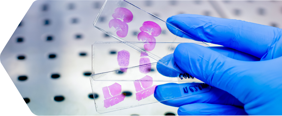
Histology Solutions
HistoSpring offers precision tissue and slide preparation, immunostaining and pathology analysis, antibody optimization, and custom requests. Our team of experts provide active collaboration and tactical solutions for successful results.
Not all staining is the same.
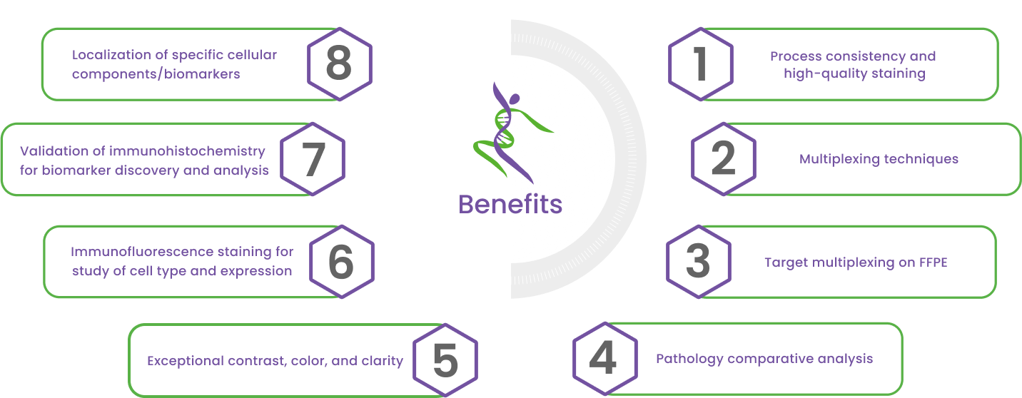
Our knowledge of chemistry, staining techniques and troubleshooting staining for tissue sections and analysis of multiple biomarkers requires expertise. Our expertise spans chemistry, staining techniques, and troubleshooting complex tissue‑based assays, including the analysis of multiple biomarkers.
Routine Histology
The path from research through pre-clinical starts with best-practice techniques, skills and sound procedures.
- Selection of fixation techniques
- Frozen OCT embedding
- Fixed tissue processing and paraffin embedding
- Microtome sectioning 4 – 20 µm
- Hematoxylin and Eosin (H & E)
Specialty Stains
HistoSpring’s team of experts provide staining techniques to enhance tissue contrast for analysis and biomarker localization.
- Alizarin Red
- In Situ Hybridization
- Masson’s Trichrome
- Multiomic Analysis (RNA + protein multiplexing)
- Naphthol-AS-D-chloroacetate Esterase
- Oil Red O
- Periodic Acid-Schiff Stain
- Picosirius Red
- Toluidine Blue
- TUNEL Assay
Immunostaining
HistoSpring offers custom antibody solutions to improve IHC results and to target cellular biomarkers and analysis. Our team of experts trouble shoot and collaborate to achieve optimum experiment results.
- Antibody optimization and validation
- Primary and secondary antibody selection and optimization
- Detection of low-abundance targets
- Fluorescent imaging

Reaching Research Targets
- Ki67
- α smooth muscle actin
- Vimentin
- Pan-Keratin
- CD3
- F4/80
- Estrogen receptor α
- Progesterone Receptor
Proliferation
- Ki-67
- PCNA
- P21
DNA /lipid damage response
- γH2AX
- 53BP1
- 8-oxo 2 deoxyguanosine
- 4HNE
- P53
Immune compartment
- CD45r
- CD3
- CD4
- CD8
- FOXP3
- CD68
- F4/80
- Arginase1
- IDO1
- Granzyme B
- PD1
- PDL1
Hormone receptors
- ER
- PR
- Neuronal markers
- GFAP
- NeuN
Cell type markers
- Cytokeratins
- Vimentin
- α smooth muscle actin
- CD31
- tdtomato