The initial development of conventional immunohistochemistry (IHC) provided the ability to examine protein expression and localization within a tissue section. For many years, however, this approach was largely limited to a single target per slide, making it difficult to understand how multiple cell types and biological signals interact within the same tissue context.
The development of multiplexed staining—using antibodies directly conjugated to different fluorophores—expanded these capabilities, though early approaches were often limited by cross-reactivity and spectral overlap. Advances in photostability, signal amplification, and spectral separation have significantly improved performance, making it possible to detect multiple proteins on the same tissue section with greater precision. This has expanded the scope of questions that can be addressed within a single thin slice of tissue.
As a result, dynamic changes in cellular composition, localization, and functional activity can now be evaluated while preserving intact tissue architecture. This approach delivers meaningful insight into both basic and translational research and is increasingly used to support personalized medicine and data-informed treatment strategies.
Why Multiplexing Adds Meaning to Spatial Immune Analysis
The value of multiplexing becomes most apparent when multiple markers are analyzed together on the same tissue section, preserving spatial immune context for interpretation. A recent review of multiplex imaging studies across several tumor types, including breast cancer, shows that spatial immune features identified through multiplex analysis are associated with response or resistance to immunotherapy¹.
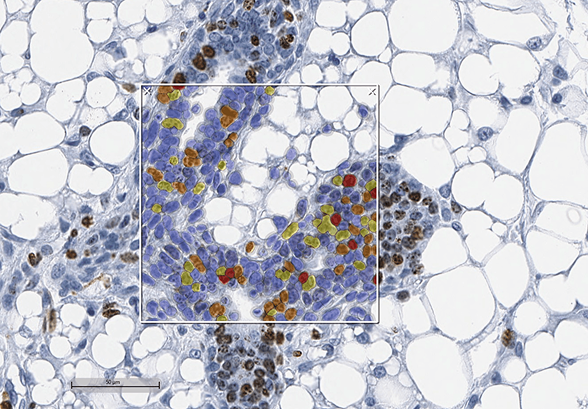
In breast cancer, multiplex analysis has shown that CD8⁺ T-cell localization, immune exclusion patterns, and the presence of tertiary lymphoid structures (TLS) correlate with therapeutic response in ways that single-marker IHC cannot capture. Multiplex imaging reveals not only which immune cells are present, but where they reside and how they are organized within the tumor microenvironment.
Tumors containing TLS or localized immune “hotspots”—enriched with activated B cells, dendritic cells, and CD8⁺ T cells—are consistently associated with more favorable immunotherapy outcomes. In contrast, immune-excluded tumors, where immune cells remain confined to tumor margins, are more likely to resist immune-based treatments.
Collectively, these findings support the investigation of how specific spatial immune patterns may influence therapeutic response and resistance, enabling more informed evaluation of therapeutic strategies.
Applying Multiplexing and Same-Section Analysis in Translational Research
As multiplexing and same-section analysis become central to modern tissue-based research, generating reliable and interpretable data depends on more than marker selection alone. Consistent staining, careful tissue handling, and robust analytical workflows are essential for translating spatial insights into meaningful biological interpretation.
HistoSpring supports multiplex immunofluorescence and advanced IHC workflows designed to preserve tissue integrity while enabling spatially resolved analysis of complex tumor microenvironments. Our team works closely with researchers to develop and execute customized marker panels that support immune profiling, functional assessment, and spatial organization analysis within tissue.
Interested in exploring multiplex IHC or same-section spatial analysis for your research?
Connect with HistoSpring to learn how we can support your work.
References
- Feng L, Li G, Zheng Y, et al. Multiplex imaging analysis of the tumor immune microenvironment for guiding precision immunotherapy. Front Immunol. 2025;16:1617906. doi:10.3389/fimmu.2025.1617906.