
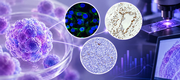
Cell models provide an important foundation for studying disease biology and therapeutic response. From established cell lines to patient-derived cultures and tissue explants, each model offers different opportunities to investigate cellular mechanisms, biological activity, and responses to experimental treatments.
But choosing the right model is only part of experimental design. The assays selected for a study are equally important. They influence what researchers can measure, which changes in cellular activity can be detected, and how findings can be interpreted.
Understanding how cell models and how assays work together can help researchers select approaches that address their scientific questions and generate meaningful data.
The Cell Model-Assay Connection: Why Both Matter
The relationship between cell models and assays goes beyond selecting the right tools for a study. The model establishes the biological context, while assays provide the means to measure cellular activity, analyze biological changes, and evaluate responses to experimental conditions.
Different assays reveal different aspects of cellular behavior. Viability and proliferation assays can assess cell survival and growth, while cytotoxicity assays measure the damaging effects of experimental treatments. Imaging, biomarker analysis, and molecular assays can provide further insight into cellular characteristics, protein expression, and signaling pathways. Combining these approaches allows researchers to examine both the effects of a treatment and the biological processes associated with those effects.
The biological characteristics of a cell model can also influence assay results. While established cell lines provide controlled systems for investigating specific mechanisms, patient-derived cultures and tissue explants can reveal responses that vary across biological settings. These differences may reflect variations in receptor expression, signaling activity, or other cellular characteristics, influencing what assays detect and how researchers interpret their findings.
Selecting the right combination helps ensure that the biological system is appropriate for the research question and that the assays provide meaningful information about the processes being investigated.
What Research Reveals About Model–Assay Combinations
Published breast research illustrates how different cell model–assay combinations can reveal individual variation, connect molecular findings with cellular activity, and identify changes that might otherwise go undetected.
Exposure to the same environmental chemical, for example, does not necessarily produce the same response across individuals. Researchers investigated the effects of PCB 126 using established cell lines, patient-derived breast epithelial cultures, and tissue explants.¹ By combining gene expression analysis with protein measurements, they identified differences among donors that highlight the importance of studying biological variation rather than relying exclusively on conventional cell lines.
Other research shows how findings from one model can lead to further investigation in another. In a study of atypical breast hyperplasia, researchers used gene expression profiling to identify changes involving SFRP1, then investigated its role using breast epithelial cell lines, mouse mammary tissue, and human breast explants.² The combination helped connect molecular findings with changes in estrogen signaling.
Even within the same tissue model, different assays can tell different parts of the story. Scientists examined estrogen responses in patient-derived breast explants and found that changes in gene expression were not consistently accompanied by changes in proliferation or apoptosis.³ Evaluating multiple endpoints revealed differences that a single measurement might have missed.
The Right Combination Makes the Difference
Selecting appropriate cell models and complementary assays can help researchers generate more informative data, strengthen the interpretation of experimental findings, and make better-informed decisions about future investigations.
Supporting Breast and Ovarian Cancer Research
HistoSpring offers breast and ovarian cell models, including patient-derived research resources, to support studies of disease biology and therapeutic response. Our experienced PhD scientists can work with researchers to identify appropriate models and assays based on their research objectives.
Contact HistoSpring to discuss your next research study.
info@histospring.com | 413-794-0523
References
- Morin SM, Majhi P, Crisi GM, et al. Interindividual variation contributes to differential PCB 126 induced gene expression in primary breast epithelial cells and tissues. Ecotoxicology and Environmental Safety. August 2022. https://www.sciencedirect.com/science/article/pii/S0147651322005620
- Gregory KJ, Roberts AL, Conlon EM, et al. Gene expression signature of atypical breast hyperplasia and regulation by SFRP1. Breast Cancer Research. 2019;21(1):76. https://pubmed.ncbi.nlm.nih.gov/31248446/
- Dunphy KA, Black A, Suresh S, et al. Inter-individual variation in response to estrogen in human breast explants. Journal of Mammary Gland Biology and Neoplasia. 2020;25(1). doi:10.1007/s10911-020-09446-3