Digitizing Histology for Deeper Tissue Insight
From whole-slide imaging to AI-powered quantitative analysis, HistoSpring delivers integrated digital pathology workflows that generate reproducible, analysis-ready data
HistoSpring’s Core Capabilities
Digital Slide Imaging & Review
High-resolution whole-slide imaging allows you to view entire tissue sections while examining cellular morphology in detail. Digital slides can be securely stored, shared, and reviewed across teams, supporting consistent interpretation.
Technology Platform
HistoSpring brings together best-in-class technologies to support digital pathology workflows—from high-resolution imaging through
quantitative analysis.
HALO® AI
A deep learning–powered image analysis platform that supports tissue classification, segmentation, and quantitative biomarker analysis, delivering consistent, reproducible, publication-ready data.
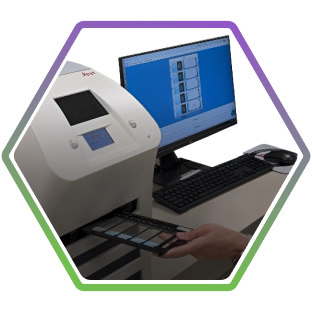
Aperio CS2
High-resolution whole-slide imaging enables digital visualization, comparison, and secure archiving of tissue sections.
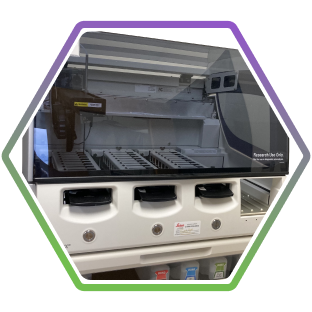
Leica BOND RXm
An automated staining platform that supports consistent, high-quality IHC, IF, and ISH workflows, generating analysis-ready slides for downstream digital analysis.
HALO® AI
A deep learning–powered image analysis platform that supports tissue classification, segmentation, and quantitative biomarker analysis, delivering
consistent, reproducible, publication-ready data.
Aperio CS2
High-resolution whole-slide imaging enables digital visualization, comparison, and secure archiving of tissue sections.
Leica BOND RXm
An automated staining platform that delivers consistent, high‑quality IHC, IF, and ISH workflows, producing slides optimized for downstream digital analysis.
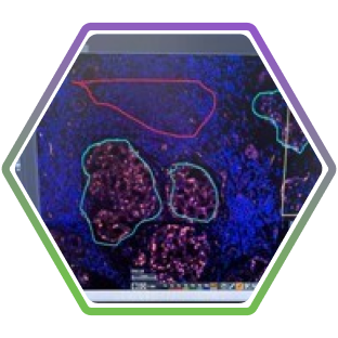
Quantitative Image Analysis & Data Generation
AI-powered image analysis enables quantitative evaluation of complex tissues with speed, consistency, and precision—transforming whole-slide images into structured, numerical data suitable for downstream interpretation.
Quantitative Analysis
- Cell-level detection and biomarker quantification
Measure expression with precision at the individual cell level - Tissue classification and segmentation
Distinguish tumor, stroma, and epithelium for context-specific analysis - Multiomic spatial analysis
Evaluate RNA and protein targets within the same section and assess spatial relationships - Whole-slide quantitative outputs
Generate cell counts, intensities, and expression levels across defined tissue compartments - Standardized, reproducible data
Enable consistent analysis across studies, cohorts, and timepoints
Custom AI Model Development
AI models can be trained and refined on specific tissue types and biomarkers to align with study-specific requirements.
- Train classifiers using annotated regions of interest
- Iteratively refine models through visual validation
- Use probability mapping to assess algorithm confidence
- Enable team review of annotations and AI output before final analysis
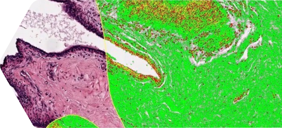
Archival Slide Re-Analysis
Previously generated slides can be re-analyzed using modern digital pathology workflows to generate new quantitative insights.
- Convert semi-quantitative scoring into quantitative data
- Expand dynamic range and improve sensitivity of detection
- Compare legacy data with current workflows
- Extract new findings without repeating wet lab experiments
How We Support Your Research
- Generate quantitative, reproducible data from complex tissue samples
- Standardize analysis across studies, timepoints, and cohorts
- Provide detailed evaluation of tissue architecture and biomarker distribution
- Support multiplex and same-section analysis for more comprehensive insight
- Reduce variability associated with manual interpretation
- Accelerate timelines from imaging through data output
Turn Your Tissue into Discovery-Ready Data
Contact us at info@histospring.com or at 413-794-0523