Where AI-Powered Precision Meets Visual, Interactive Science
From Click-Counting to Command Center
“HALO AI is the new video game for pathologists.” That’s how one researcher described it—and for good reason. Histology labs are rapidly evolving from manual click-counting to intelligent, AI-driven image analysis. Today, scientists don’t just review stained slides—they interact with them.
HALO transforms traditional digital pathology into an interactive, high-performance experience. With game-like visualization and real-time feedback, it replaces subjective counts with fast, reproducible data that bring confidence to every study.
What It Is—and Why It Feels Like Play
HALO-AI is a deep learning-powered image analysis platform designed to modernize tissue interpretation. It allows researchers to train models on specific tissue types, set detection parameters, and validate results—all in a highly visual, intuitive environment.
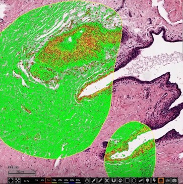
Through real-time overlays, color-coded masks, and iterative model refinement, researchers can fine-tune their analyses much like mastering a video game level. The result? A platform that’s not only efficient but immersive and scientifically precise.
The Serious Power Behind the Interface
Beyond its intuitive interface, this advanced platform revolutionizes tissue image analysis by turning labor-intensive manual counting into rapid, quantitative workflows. By removing human subjectivity and delivering consistent, algorithm-driven assessments, HALO-AI helps researchers achieve both speed and reliability in their data.
Key advantages include:
- Speed: Analyses that once took hours now finish in minutes.
- Consistency: Every slide is assessed with the same trained model, eliminating variability.
- Confidence: Visual validation through annotated images before final outputs are generated.
- Collaboration: Overlays and annotations can be easily reviewed with colleagues, ensuring alignment before data export.
Transforming Tissue-Based Research
The platform supports a wide range of applications—from studying tumor microenvironments and immune cell infiltration to analyzing tissue and cell structure. Researchers gain standardized, reproducible insights that move beyond qualitative impressions to robust, quantitative data.
Key focus areas include:
- Biomarker expression analysis
- Immune cell density and distribution
- Tumor heterogeneity and morphology
AI Insights: Driving Research Forward
AI is making a measurable impact on histology and pathology, delivering unmatched precision and reproducibility.
The esophageal squamous cell carcinoma study¹ demonstrated how a deep learning classifier was used to quantify tumor cell nuclear size (NS) and link it to patient outcomes. Larger nuclei were associated with poorer survival, establishing NS as a new prognostic marker and highlighting how AI can uncover subtle features that inform clinical decisions.
In a separate study focused on Sjogren’s Disease², a custom-trained tissue classifier accurately identified and quantified stroma, glandular tissue, adipose tissue, and immune infiltrates in salivary gland samples—delivering detailed, reproducible data in seconds. This enables rapid, standardized evaluations and offers new clarity on disease mechanisms and progression, all without the manual workload.
In a separate line of research, prostate cancer studies confirmed that an AI-driven algorithm can match expert pathologists in cancer detection and Gleason grading³—even identifying tumor areas initially missed during human review. This supports more consistent, accurate diagnoses and helps improve treatment decision-making across diverse clinical settings.
Together, these studies highlight how AI-powered image analysis is driving forward discoveries, offering deeper insights that were previously out of reach.
Conclusion: Precision Meets Play
Advanced AI tools are transforming digital pathology from a static review process into an interactive, data-driven experience. Researchers now have intelligent ways to quantify complex tissues, validate interpretations collaboratively, and gain consistent, reproducible insights—without the burden of manual analysis.
It’s not just a technological upgrade—it’s a meaningful leap forward in how labs can deliver confidence, clarity, and scientific excellence to every tissue study.
Ready to bring next-level image analysis to your research?
HistoSpring now offers HALO AI image quantification as part of our expanded histology services. With this advanced technology, we can:
- Accurately annotate your regions of interest
- Collaborate closely to validate interpretations before analysis
- Quantify complex features using custom-trained AI algorithms
- Deliver detailed, spreadsheet-ready data with visual confirmation
Learn more about this exciting new technology. Contact us at histololgy@histopsring.com or at 413-794-094.
References
- Keita Kouzu, Hironori Tsujimoto, Ines P. Nearchou, et al. Prognostic Impact of Tumor Cell Nuclear Size Assessed by Artificial Intelligence in Esophageal Squamous Cell Carcinoma. Laboratory Investigation.
March 2025. https://www.sciencedirect.com/science/article/abs/pii/S0023683724018993 - M. C. Marlin, L. Bradshaw, K. Wright, et al. AB0802 AI-Enabled Tissue Classifier for Sjogren’s Disease Salivary Gland Identifies Key Histological Features Important for Understanding Disease Manifestation. Annals of Rheumatic Diseases. June 2024. https://ard.bmj.com/content/83/Suppl_1/1695.1
- Yuri Tolkach, Vlado Ovtcharov, Alexey Pryalukhin, et al. An International Multi-Institutional Validation Study of the Algorithm for Prostate Cancer Detection and Gleason Grading. NPJ Precision Oncology. August 2023. https://www.nature.com/articles/s41698-023-00424-6
#DigitalPathology #SpatialBiology #PrecisionMedicine #CancerResearch