
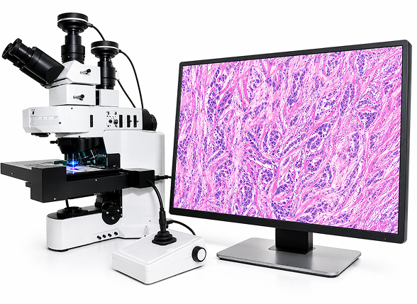
As molecular technologies continue to advance, the ability to isolate specific cell populations from heterogeneous tissues has become increasingly important. Genomics, transcriptomics, proteomics, and spatial biology continue to advance biological research, but the quality and biological relevance of these analyses depend on accurately isolating the cells being studied.
Laser Capture Microdissection (LCM) enables researchers to precisely isolate targeted cell populations while preserving their molecular integrity for downstream analysis. Integrated with histology, immunohistochemistry, and digital pathology, LCM helps decipher complex tissue and support more precise molecular characterization, biomarker discovery, and advanced tissue research.
Enabling Precise Cell Isolation Through Histology and LCM
Histology provides the morphological framework for identifying specific cell populations within heterogeneous tissues prior to laser capture microdissection. By preserving tissue architecture, histological evaluation enables researchers to select targeted regions for precise cell isolation, providing the foundation for accurate downstream molecular analysis.
By minimizing contamination from surrounding cells, LCM improves the accuracy of downstream molecular analyses and supports a broad range of applications, including biomarker discovery, gene expression profiling, RNA sequencing, spatial transcriptomics, and precision medicine research.
In one study of atypical breast hyperplasia, researchers used histology-guided laser capture microdissection to isolate histologically normal and atypical epithelial tissues for transcriptomic analysis. Paired analysis of histologically normal and atypical hyperplasia tissues identified a 99-gene expression signature that discriminated atypical hyperplasia from histologically normal breast tissue.¹ Together, histology and laser capture microdissection enable researchers to isolate specific cell populations for more precise molecular characterization of complex tissues.
Advancing Biomarker Discovery Through Precise Cell Isolation
Biomarker discovery depends on accurately characterizing the molecular changes that drive disease. In heterogeneous tissues, analyzing mixed cell populations can obscure clinically relevant molecular signals, making it more difficult to identify biomarkers associated with disease progression, therapeutic response, and patient stratification.
By enabling precise cell isolation from heterogeneous tissues, LCM helps researchers identify disease-associated biomarkers with greater confidence, improving molecular characterization and strengthening studies of disease progression, therapeutic response, and precision medicine.
A subsequent breast cancer study further demonstrated the value of laser capture microdissection for molecular characterization through genome-wide DNA methylation analysis. Researchers used laser capture microdissection to isolate epithelial cells from breast tumor tissue and matched histologically normal tissue before profiling DNA methylation. The study identified DNA methylation alterations consistent with breast field cancerization and concluded that these findings may provide an opportunity to develop epigenetic biomarkers of disease risk.²
Together, these capabilities make laser capture microdissection a valuable tool for biomarker discovery and molecular characterization.
Integrating LCM with Digital Pathology
Integrating digital pathology with laser capture microdissection creates a more standardized and reproducible approach to tissue selection by combining whole-slide imaging, digital image analysis, AI-assisted tissue annotation, and precise cell isolation. Automated region selection helps reduce observer variability, improve workflow efficiency, and enable researchers to consistently isolate the most biologically relevant cell populations from complex tissue specimens. Together, these technologies support more scalable molecular workflows while improving the quality and consistency of downstream analyses.
Advances in digital pathology are expanding the capabilities of laser capture microdissection by integrating automated image analysis with tissue selection workflows. These emerging approaches support more efficient, scalable spatial molecular analysis of FFPE tissue specimens, creating new opportunities for biomarker discovery and translational research.³
The Growing Importance of Laser Capture Microdissection
As molecular technologies continue to evolve, precise cell isolation has become increasingly important for generating high-quality molecular data and advancing translational research. Integrated with histology, immunohistochemistry, biomarker analysis, and digital pathology, laser capture microdissection enables researchers to generate more accurate, cell-specific molecular insights for advanced tissue research.
HistoSpring combines advanced laser capture microdissection technology with integrated histology and digital pathology services to deliver customized workflows that support advanced tissue research. Contact us to learn how we can support your next research project, info@histospring.com | 413-794-0523.
References
- Gregory KJ, Roberts AL, Conlon EM, et al. Gene expression signature of atypical breast hyperplasia and regulation by SFRP1. Breast Cancer Research. 2019;21(1):76. doi:10.1186/s13058-019-1157-5.
- Muse ME, Titus AJ, Salas LA, et al. Enrichment of CpG island shore region hypermethylation in epigenetic breast field cancerization. Epigenetics. 2020;15(10):1093-1106. doi:10.1080/15592294.2020.1747748.
- Gasparello J, Pearson RC, von Kriegsheim A. Artificial intelligence-driven laser capture microdissection. Methods in Molecular Biology. 2026;3015. doi:10.1007/978-1-0716-4500-8_7.