At the beginning of this two-part blog series, we focused on the critical components of wet lab spaces by detailing commonly required tools to drive research in the life sciences — real-time PCR machines, plate readers, and biological irradiators.
Tissue Culture Facilities
Equipping a fully functional tissue culture lab can be a financial hurdle for most startups. HistoSpring mitigates this burden for its research partners by sharing full facility access to its robust tissue culture facility located at Baystate Research Facility. Baystate’s lab contains all necessary equipment including Biological Safety Cabinets (BSCs), specifically the SterilGARD and SterilGARD III Advance models.
SterilGARD BSCs offer a sterile work environment to safeguard cell cultures from contaminants, ensuring experiments’ accuracy and reliability — a critical factor for researchers seeking successful results.
SterilGARD BSCs include tailored features for efficient research:
- Ergonomic Design: An angled work surface promotes a comfortable posture, reducing fatigue during extended periods of cell manipulation.
- Easy Maintenance and Cleaning: Smooth, coved interior surfaces facilitate thorough cleaning and disinfection, minimizing downtime between experiments.
- Advanced HEPA Filtration: Both of these BSCs utilize High-Efficiency Particulate Air (HEPA) filters. These filters remove over 99.97% of airborne particles larger than 0.3 microns, protecting the cell cultures and researchers from harmful microorganisms. This enables aseptic manipulation of cells for various applications, including:
- Media changes
- Transfections
- Experiment set-up
With six hoods, including three SterilGUARD III Advance models, biotech startups are able to conduct research with greater efficiency and affordability.
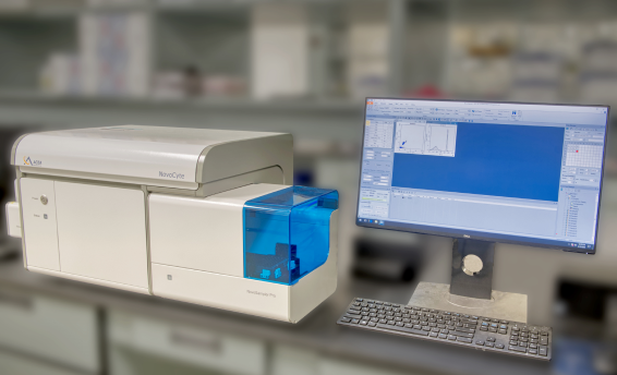
Flow Cytometry and Biomarker Detection
A flexible tool for biomarker detection is the use of flow cytometry to analyze specific characteristics that indicate normal cellular function versus disease progression. Flow cytometry offers researchers multiple benefits, including:
- Multiparameter Analysis: Researchers can use this tool to simultaneously analyze multiple cellular characteristics, such as size, granularity, and the presence of specific proteins.
- Single Cell Analysis: Enables examining individual cells within a population, providing valuable insights into cellular heterogeneity and potential drug response variations.
- Cell Function Determination: Investigators can use flow cytometry for immunophenotyping assays, cell cycle analysis, and apoptosis studies. These types of experiments are crucial for comprehensive studies in immunology, cancer research, and beyond.
Researchers have access to the NovoExpress with 1.6.2 software compliant with 21 CFR Part 11, ensuring data integrity and adherence to regulatory requirements.
Advanced Microscopy
HistoSpring equips researchers with not just one but two highly functional microscopes: the Nikon TE2000 and the Nikon Eclipse Ti Fluorescent Microscope.
These Nikons play a vital role in visualizing and understanding the intricate world of cells, opening doors for breakthroughs in many areas of life sciences.
Fluorescent Microscopy
The TE2000 and the Eclipse Ti serve as foundational instruments offering researchers:
- High-quality brightfield and DIC imaging: This allows for clear visualization of cellular morphology, providing a detailed picture of cell size, shape, and internal structures.
- Versatility for diverse applications: Both microscopes can adapt for various techniques like phase contrast and differential interference contrast (DIC).
- Advanced Fluorescence Imaging: The TE2000 and the Ti are designed to observe fluorescently labeled cells. Fluorescent proteins allow researchers to target and monitor specific cellular components, providing deeper insights into cellular processes.
- Ideal for Cancer Research: Cancer cells often exhibit distinct fluorescent properties. Advanced imaging capabilities empower researchers to study these features, aiding in cancer diagnosis, treatment development, and monitoring treatment response.
One Step Further
The Eclipse Ti takes cellular visualization to the next level by offering all of the capabilities listed above by adding live cell imaging.
- Live Cell Imaging: The Nikon Eclipse Ti Fluorescent microscope can facilitate dynamic, real-time observations of living cells. The non-invasive nature of fluorescence imaging allows researchers to track cellular events over time without disrupting the natural physiological state of the cells. Live cell imaging is invaluable for studying dynamic processes, including cell migration, division, and responses to external stimuli, and it allows us to advance our understanding of living systems.
The complementary approach of these microscopes is invaluable in research, specifically in cancer research, as fluorescence imaging and bioluminescence imaging are being used to identify tumors for staging and tracking novel therapeutic agents.1
Resources to Unleash Research
Limited resources should never hinder research potential. HistoSpring’s state-of-the-art facilities and collaborative environment provide the perfect launchpad for various research projects.
Learn more about HistoSpring’s research space and advanced tools — fill out this form to get started!
References
- Huang S, Jiang C, Mughal MJ, Wang G, Xing F. An In Vivo Fluorescence Resonance Energy Transfer-Based Imaging Platform for Targeted Drug Discovery and Cancer Therapy. Frontiers in Bioengineering and Biotechnology. 2022;10. doi:https://doi.org/10.3389/fbioe.2022.839078